The patient had no dyspnea or chest tightness. No other symptoms such as cough, nausea, vomiting or fever were reported. Although a musculoskeletal etiology was suspected by the patient, there was no trauma or precipitating event. She presented to exclude pulmonary embolism, given a positive family history. The patient was not taking any regular medication or hormonal contraception.
On clinical examination, pain was not provoked by manual pressure or physical movement. The patient was hemodynamically stable (HR 68 bpm, BP 96/62 mmHg) and mildly tachypneic (23/min) with adequate saturation (SpO2 95%). Blood tests showed normal leukocytes (9.7 G/l) with normal hemoglobin and slightly elevated D-dimer (607 ng FEU/ml). ECG and cardiac biomarkers were normal.
A chest CT scan with intravenous contrast was performed to rule out a pulmonary embolism and revealed the following image:
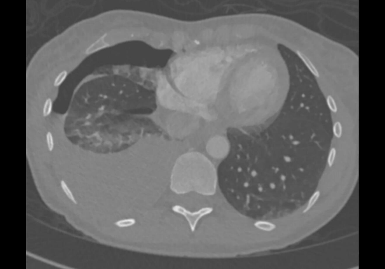
Figure 1: Chest CT scan with intravenous contrast.
Question 1: A mean pleural effusion density of 38 Hounsfield units (HU) was measured on CT. What is the most likely type of pleural effusion?
A: Exudative effusion
B: Hemothorax with a largely fluid component
C: Transudative effusion
D: Hemothorax with a largely clotted component
The Hounsfield scale describes the radiodensity in medical CT. Pure H2O is defined as 0 HU. CT is not a sensitive tool for differentiating between transudates and exudates. However, exudates have higher median HU values (12.5 HU exudate vs. 5 HU transudate).1 Fluid blood has HU values between 30 and 45. Clotted blood has a high attenuation of 45 to 70 HU.2 In the presented case, a hemopneumothorax with a largely fluid component was diagnosed based on the CT findings.
A chest tube (Bülau) was placed. Immediately after placement, 1 liter of blood was drained. The patient remained hemodynamically stable despite a drop in hemoglobin and was transferred to intensive care for monitoring. Transfer to a tertiary care center for video-assisted thoracoscopic surgery (VATS) was arranged due to retained hemothorax on control x-ray after chest tube placement.
The following findings were observed during VATS:
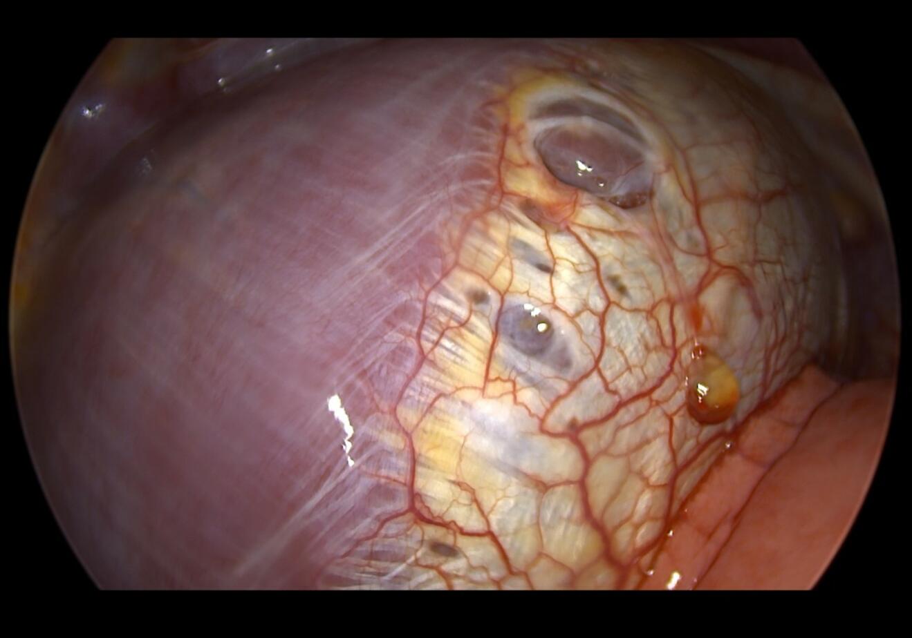
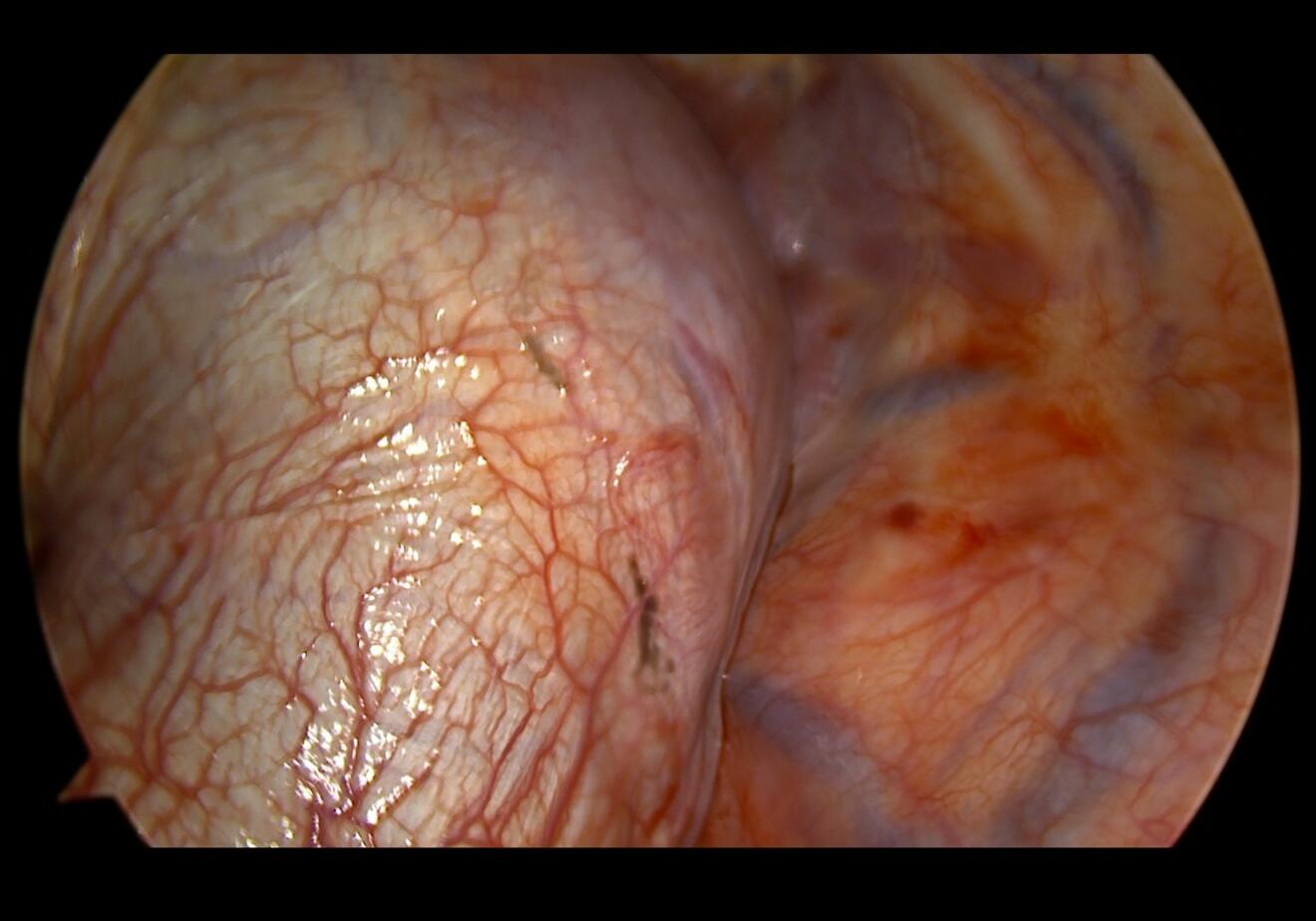
Figure 2: Intraoperative findings VATS, diaphragm visualized.
The images show changes in the centrum tendineum of the diaphragm. The parietal and visceral pleura showed no abnormalities. The lesions were biopsied for histological analysis and the defects were closed. Histological analysis revealed connective tissue with anthracosis and histiocytic reaction, and a single gland positive for estrogen receptor.
Question 2: Based on the findings, what is the correct diagnosis?
A: Thoracic wall malignancy
B: Connective tissue disorder, Ehlers-Danlos
C: Lung malignancy
D: Endometriosis
E: Hepatocellular malignancy with diaphragmatic infiltration
This case describes thoracic endometriosis syndrome (TES) presenting as a massive catamenial hemopneumothorax. Catamenial is derived from the Greek word "katamenios", meaning monthly occurrence.
Endometriosis is defined as the presence of endometrium-like epithelium and/or stroma outside of the uterus, affecting roughly 10% of all women of reproductive age.3 The disease is most commonly diagnosed in the abdominopelvic cavity and can have a significant impact on the patient's quality of life. Extra-abdominal manifestations are known, thoracic endometriosis being the most common site outside the abdominopelvic cavity. TES has a later onset than abdominopelvic endometriosis, most often coexists with it and is therefore considered to be a progression of the disease.4
As the lung parenchyma, pleural surfaces and/or diaphragm may be pathologically involved, TES encompasses a range of clinical symptoms that correlate with the anatomical location of the lesions. Catamenial pneumothorax is the most common manifestation of the disease and hemopneumothorax is seen in a minority of cases.5
TES is nine times more common in the right thoracic cavity than in the left. Although the pathophysiology is not fully understood, this observation supports Sampson's theory of retrograde menstruation. Sampson's theory suggests that endometrial cells enter the peritoneal cavity via the fallopian tubes and follow the physiological right paracolic flow to the right hemidiaphragm. The flow is redirected away from the left hemidiaphragm by the falciform and phrenicocolic ligaments. After seeding, the endometrial cells are thought to migrate into the thoracic cavity.4
VATS is considered the gold standard for diagnosis and treatment of TES. Several techniques can be used to inactivate or excise endometriotic lesions in a parenchyma-sparing manner. Pleurodesis has a role in preventing future recurrence of pneumothorax. As abdominopelvic endometriosis is suspected in the context of TES, especially in the presence of diaphragmatic lesions, the literature suggests a routine role for diagnostic laparoscopy combined with VATS. Medical treatment consists of combined hormonal contraceptives, progestogens, GnRH agonists or GnRH antagonists, and patients should be counseled by a specialist gynecologist.4
The early postoperative course of this patient was uneventful and she was discharged on the second postoperative day after gynecological consultation with initiation of hormone therapy (Dienogest). Although the available literature suggests a role for proactive search for abdominopelvic endometriosis, we believe that invasive diagnostic and treatment decisions should be based on individual assessment. Our patient did not have a clear history of dysmenorrhea and in line with the patient's preferences we decided against a diagnostic laparoscopy.
Approximately 1.5 months after the initial diagnosis, the patient presented again with a right-sided catamenial pneumothorax. Re-VATS revealed a new endometriotic lesion on the visceral pleura of the right upper lobe. A small wedge resection with talc pleurodesis was successfully performed.
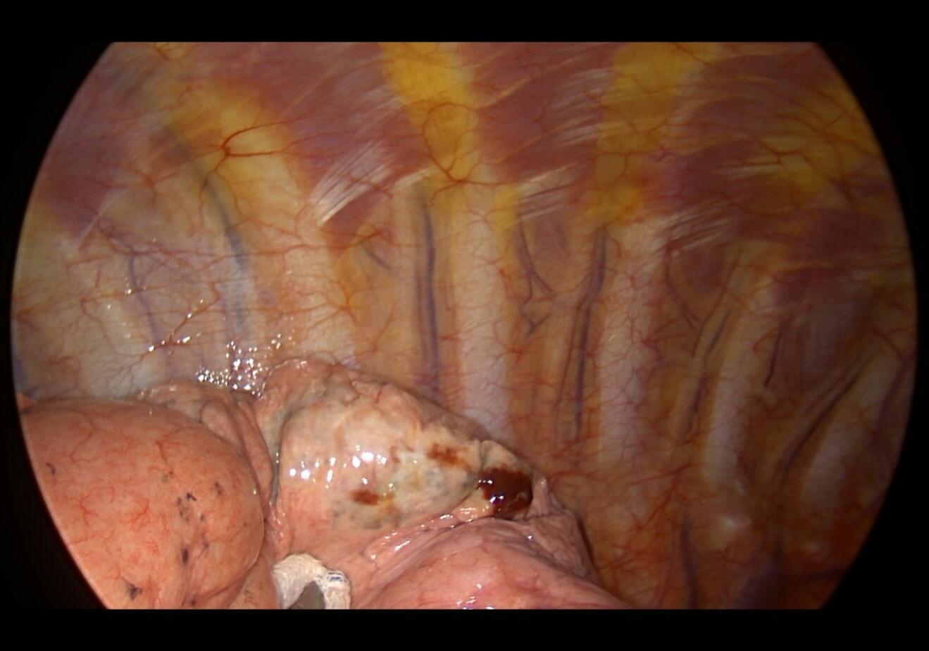
Figure 3: Intraoperative findings Re-VATS, right upper lobe visualized.
1. Çullu N, Kalemci S, Karakaş Ö, Eser İ, Yalçin F, Boyacı FN, Karakaş E. Efficacy of CT in diagnosis of transudates and exudates in patients with pleural effusion. Diagn Interv Radiol. 2014 Mar-Apr;20(2):116-20. doi: 10.5152/dir.2013.13066. PMID: 24100060; PMCID: PMC4463296.
2. DenOtter TD, Schubert J. Hounsfield Unit. 2023 Mar 6. In: StatPearls [Internet]. Treasure Island (FL): StatPearls Publishing; 2024 Jan–. PMID: 31613501.
3. World Health Organization. Endometriosis. Fact sheet, 24 March 2023.
4. Nezhat C, Lindheim SR, Backhus L, Vu M, Vang N, Nezhat A, Nezhat C. Thoracic Endometriosis Syndrome: A Review of Diagnosis and Management. JSLS. 2019 Jul-Sep;23(3):e2019.00029. doi: 10.4293/JSLS.2019.00029. PMID: 31427853; PMCID: PMC6684338.
5. Agossou M, Sanchez BG, Alauzen PH, Olivier M, Cécilia-Joseph E, Chevallier L, Jean-Laurent M, Aline-Fardin A, Dramé M, Venissac N. Thoracic Endometriosis Syndrome (TES) in Martinique, a French West Indies Island. J Clin Med. 2023 Aug 26;12(17):5578. doi: 10.3390/jcm12175578. PMID: 37685644; PMCID: PMC10488738.