

Case Description
A 24-year-old male presented to the emergency department with severe lower abdominal pain. The symptoms had begun the previous day, initially improved overnight, and then recurred with marked worsening on the day of presentation. Nausea, vomiting, fever, and any changes in bowel or urinary habits were denied. The patient had no prior surgical history. His regular medication consisted only of quetiapine, which had recently been initiated for insomnia associated with a depressive episode. He reported regular use of cannabis, amphetamines, and alcohol.
On presentation, the patient was hemodynamically stable and had normal vital signs. Physical examination revealed mild scleral icterus, which was also known from the patient’s brother. The abdomen was soft, with tenderness in the right hemiabdomen and p. m. in the right lower quadrant.
Laboratory investigations were unremarkable except for an elevated total bilirubin level of 34 µmol/l.
Abdominal ultrasonography showed no pathological findings. Subsequent abdominal CT imaging demonstrated a medialized, stool-filled cecum with marked dilatation and a twisted vascular pedicle (Figure A).
Due to progressive clinical deterioration, characterized by severe abdominal pain and the development of localized peritoneal signs the patient ultimately underwent diagnostic laparoscopy.
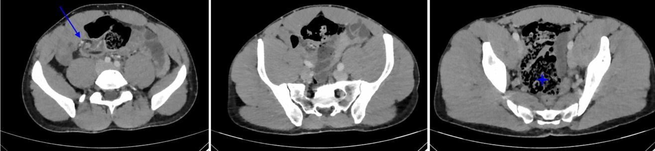
Figure A: Computed tomography demonstrating the twisted vascular pedicle (blue arrow) and a medialized cecum (blue star)
Question
What is the most likely intraoperative finding?
A. Acute gangrenous appendicitis with perforation
B. Internal herniation of the small bowel through a mesenteric defect
C. Meckel’s diverticulitis with localized peritonitis
D. Axial ileocolic cecal volvulus with a mobile cecum
E. Sigmoid volvulus causing large bowel obstruction
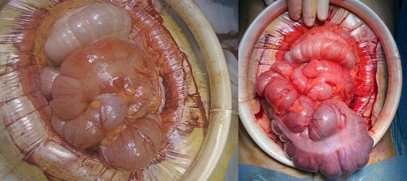
Figure B: Intraoperative pictures of the the cecal volvulus on the left side; on the right side after detorsion.
Case Solution
Intraoperatively, a cecal volvulus was identified. Due to poor visualization, the procedure was converted to an open laparotomy. Exploration revealed an axial ileocolic cecal volvulus, which was completely detorsed with two counterclockwise rotations (Figure B). A mobile cecum was subsequently noted. As the bowel was viable and there was no evidence of intestinal malrotation, an ileocecal resection with side-to-side ileoascendostomy was performed.
The elevated bilirubin level with associated scleral icterus was considered consistent with Gilbert’s syndrome. The hyperbilirubinemia was presumed to have been triggered by physiological stress related to insomnia and depression. The postoperative course was uneventful, and the patient was discharged home a few days later.
The exact prevalence of a mobile cecum in the general population remains unknown, as the condition is usually asymptomatic and typically detected only when complications occur, as illustrated by the present case. It represents an anatomical variant caused by incomplete embryologic fixation of the cecum and ascending colon to the lateral peritoneum.[1] This results in hypermobility and predisposes affected individuals to colonic torsion around its mesentery.
Cecal volvulus most commonly presents with non-specific and acutely developing symptoms such as abdominal pain. Additional possible symptoms include abdominal distension, nausea and vomiting, constipation, and bloating. Cecal volvulus typically presents in younger patients and has a female predominance.[2], [3]
Characteristic radiologic findings include marked cecal dilatation and, in some cases, an ectopic position of the cecum. A dilated, air-filled cecum may produce the so-called coffee-bean sign. The whirl sign, representing twisting of the mesenteric vessels and mesentery around the point of torsion, is considered the most specific CT finding for cecal volvulus, particularly when observed in conjunction with a markedly dilated and displaced cecum. In our case however, it was the twisted vascular pedicle.
Early diagnosis and prompt surgical intervention are essential to prevent bowel ischemia and perforation. Segmental resection with primary anastomosis is generally considered the treatment of choice when bowel viability permits. Resection is favoured because it is associated with the lowest recurrence rate. Non-resectional procedures, such as cecopexy or detorsion alone, should be limited to patients who are not suitable candidates for resection.2
[1] Mobile Cecum Syndrome. Diseases of the Colon and Rectum. 1984. Rogers RL, Harford FJ.
[2] The American Society of Colon and Rectal Surgeons Clinical Practice Guidelines for the Management of Colonic Volvulus and Acute Colonic Pseudo-Obstruction. Diseases of the Colon and Rectum. 2021. Alavi K, Poylin V, Davids JS, et al.
[3] Gastrointestinal Surgical Emergencies Textbook. American College of Surgeons (2021). 2020. Ashley E. Aaron, Andrea Amabile, Ciro Andolfi, et al.